Aim of this work was to evaluate the efficacy of a new antibiotic bone substitute, Calcium Sulphate Hemihydrate + Hydroxyapatite + Gentamicin Sulfate (CSH + HA + GS), in the treatment of osteomyelitis (OM) in diabetic foot.
Methods
From June 2013 to February 2016 we used a new CSH + HA + GS compound to fill resected bone voids following surgical intervention in cases of diabetic foot OM. The uniqueness of this product is that it induces native bone growth, while the synthetic bone disappears and antibiotic is released into the surrounding tissues, maintaining high gentamicin concentrations for some weeks.
CeramentTM |G
In 24 patients, with (8, 33,3%) or without (16, 66,7%) Charcot neuroarthropathy and post-lesional osteomyelitis, after removal of infected bone we applied 10 to 20 ml CSH + HA + GS, filling the residual spaces and aiming to stabilize the remaining bone fragments. When needed, these arthrodeses were stabilized by external-internal hybrid fixators. X-ray evaluations and, when indicated, MRI evaluations were performed before and after surgical intervention, and 3 months post-op. Revascularization with percutaneous angioplasty was performed when needed (13/24, 54.1%). 24 patients affected by OM were treated, 4 of them having 1st metatarsal head involvement, 7 having heel involvement, 13 tarsal and hindfoot involvement (Table 1). After surgical intervention all of them were treated with standard medication and pressure relief.
Place of lesion
No. of patients
Metatarsal head involvement
4
Heel involvement
7
Tarsal and hindfoot involvement
13
Table 1 – No. of patients and place of lesion
Results
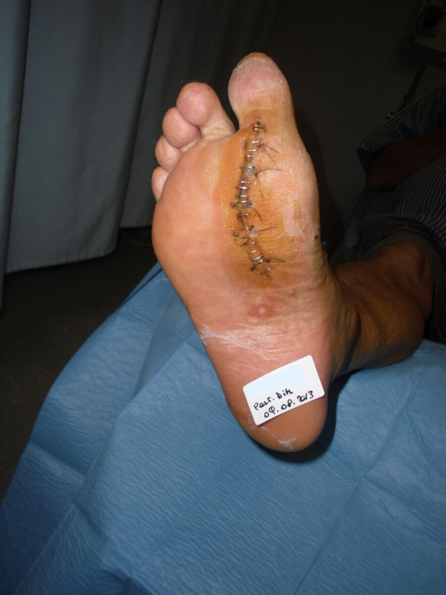
The four 1st metatarsal OM cases healed (100%), both in regards to infection and lesions (Figure 1).
First metatarsal diabetic foot ulcer (preoperative)
Preoperative X-ray
Post operative diabetic foot
Postoperative X-ray
Figure 1
One (14,3%) of the patients with heel OM presented with a worsening of the infection and was treated by major amputation, one patient (14,3%) died during early follow-up, due to cardiovascular causes; all the remaining five patients (71,4%) healed, one of them was presenting good soft tissue growth two months from the intervention, and in the absence of clinical signs of OM relapse, was treated with a sural fasciocutaneous pedicled flap.
Nine (69,2%) of the 13 patients who had midfoot or hindfoot partial resections healed (Figure 2), one patient is still ongoing, 3 (23%) patients presented late OM relapse or lesions evolution, treated with minor amputations.
Preoperative diabetic foot
Preoperative diabetic foot
Preoperative X-ray
Preoperative X-ray
Arthrodesis with external hybrid fixator
Arthrodesis with external hybrid fixator
Arthrodesis with external hybrid fixator
Postoperative X-ray
Postoperative X-ray
Healed foot
Healed foot
Figure 2
The healed patients are all wearing suitable shoes (Figure 3).
Appropriate custom-made footwear
Appropriate custom-made footwear
Figure 3
Conclusion
The use of a new CSH + HA + GS bone substitute has shown to be efficacious in inducing OM healing and preserving foot structures in diabetic feet.
Multidisciplinary approach for the management and… Diabetic patients are at increased risk of developing foot ulcers which may cause bone infections associated with a high probability of both amputation and mortality. Therefore, prompt diagnosis and adequate treatment are of key importance. In our Diabetic Foot Unit, effective multidisciplinary treatment of osteomyelitis secondary to diabetes involves the application of a gentamicin-eluting calcium…
Radiological and clinical outcomes in the medium… The aim of this work was to evaluate, via foot and ankle MRI, the outcomes of the use of a bone substitute (CERAMENT™|G) and the growth of native bone in the treatment of osteomyelitis (OM) of the diabetic foot. In nine patients from July 2014 to December 2016 we used a Calcium Sulphate Hemihydrate +…
Curriculum I am Head of the Diabetic Foot Unit of the Abano Polyclinic (Abano Terme, Padua, Italy) and I have been in this position since the beginning of 2023. Since 2010 I have been part of the team taking care of patients in the ward, treating outpatients and performing surgery (wound treatment, limb salvage, reconstructive surgery,…
Dr Christine Whisstock
Medical doctorand endocrinologist, specialised in the treatment of diabetes and of the diabetic foot.
PhD in hypertension and prevention of cardiovascular risk.
We ask your consent to use cookies to collect statistics in order to improve your browsing experience and the contents of the website. Our suppliers will receive information on how you use our website. You can change your preferences at any time by opening this panel located at the bottom left of the browser window. Learn more: Cookies Policy
Functional Always active
We use technical / functional cookies to provide our service and for security reasons. Technical cookies do not provide user profiling and cannot be disabled.
Preferences
L'archiviazione tecnica o l'accesso sono necessari per lo scopo legittimo di memorizzare le preferenze che non sono richieste dall'abbonato o dall'utente.
Statistics
We use third-party statistical cookies to track user visits to the website. This allows us to know how many people visit the website and how they interact with the contents in order to improve them.L'archiviazione tecnica o l'accesso che viene utilizzato esclusivamente per scopi statistici anonimi. Senza un mandato di comparizione, una conformità volontaria da parte del vostro Fornitore di Servizi Internet, o ulteriori registrazioni da parte di terzi, le informazioni memorizzate o recuperate per questo scopo da sole non possono di solito essere utilizzate per l'identificazione.
Marketing
We use third-party marketing cookies to be able to provide their services (Google Maps, Google YouTube, etc.). The list of suppliers and their Privacy Policy can be found in the website's Cookie Policy.